


Diagnostics 2024, 14(9), 922; https://doi.org/10.3390/diagnostics14090922 (registering DOI) - 29 Apr 2024
Abstract
Background: The polymerase chain reaction of upper respiratory tract swab samples was established as the gold standard procedure for diagnosing SARS-CoV-2 during the COVID pandemic. However, saliva collection has attracted attention as an alternative diagnostic collection method. The goal of this study was
[...] Read more.
Background: The polymerase chain reaction of upper respiratory tract swab samples was established as the gold standard procedure for diagnosing SARS-CoV-2 during the COVID pandemic. However, saliva collection has attracted attention as an alternative diagnostic collection method. The goal of this study was to compare the use of saliva and nasopharyngeal swab (NPS) samples for the detection of SARS-CoV-2. Methods: Ninety-nine paired samples were evaluated for the detection of SARS-CoV-2 by saliva and swab for a qualitative diagnosis and quantitative comparison of viral particles. Furthermore, the detection limits for each sample collection technique were determined. The cycle threshold (CT) values of the saliva samples, the vaccination status, and the financial costs associated with each collection technique were compared. Results: The results showed qualitative equivalence in diagnosis (96.96%) comparing saliva and swab collection, although there was low quantitative agreement. Furthermore, the detection limit test demonstrated equivalence for both collection methods. We did not observe a statistically significant association between CT values and vaccination status, indicating that the vaccine had no influence on viral load at diagnosis. Finally, we observed that the use of saliva incurs lower financial costs and requires less use of plastic materials, making it more sustainable. Conclusions: These findings support the adoption of saliva collection as a feasible and sustainable alternative to the diagnosis of COVID-19.
Full article
(This article belongs to the Section Diagnostic Microbiology and Infectious Disease)
►
Show Figures
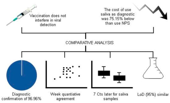
Figure 1

